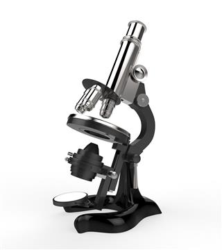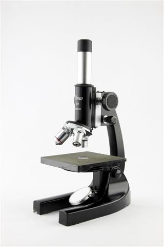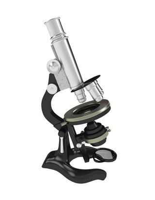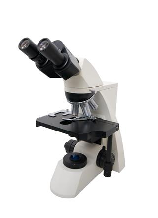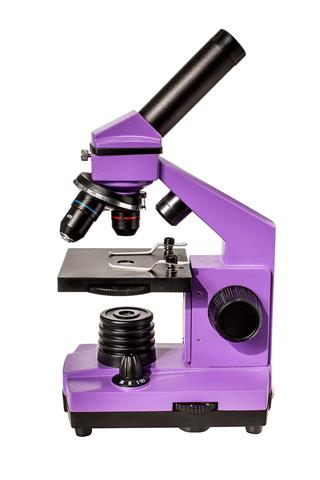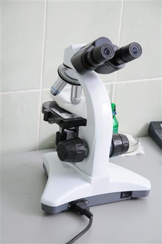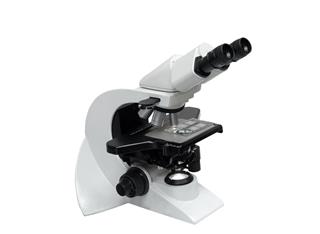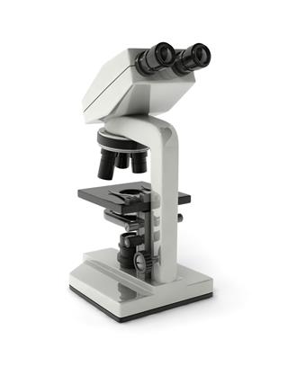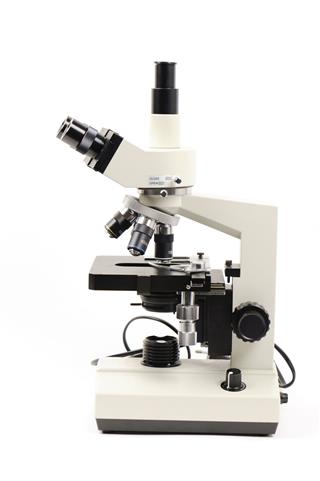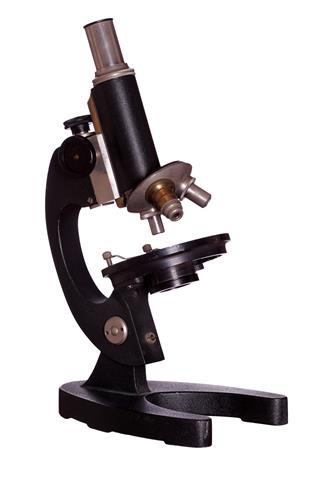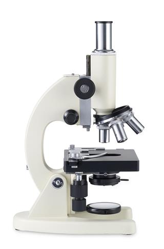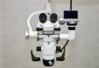
Compound microscope is a widely used instrument in the field of life sciences helps solve many mysteries of life. The following article will cover information on its parts and functions.
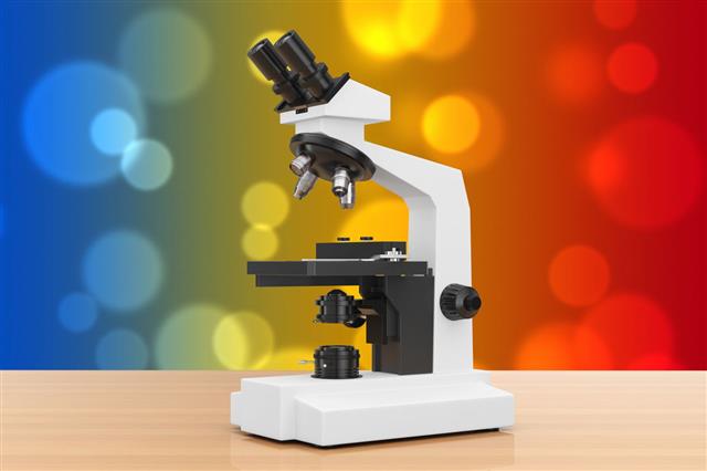
A compound microscopes helps in magnifying an image in two stages. It uses an objective lens that has many powers on a turret and an eyepiece that helps in magnifying the image formed by the objective lens. It is divided into structural parts and optical parts.
Labeled Diagram of a Compound Microscope

Structural Parts
It is divided into three basic structural components, which can be explained as follows:
Head:
The head or body of a compound microscope contains the optical parts of the microscope.
Base:
The base of a compound microscope is helps in supporting the microscope and contains the illuminator.
Arm:
The arm acts as a connector between the base and the head of the compound microscope.
Optical Parts
There are various optical parts of a microscope that help one observe the specimen or samples on a slide.
Eyepiece:
The eyepiece is the ocular lens that helps you look through to see a magnified image from the top of the microscope. The lens have a power of magnification of about 10x or 15x.
Eyepiece Tube:
The part that connects the eyepiece with the objective lens is the tube.
Turret:
The nose piece that supports the objective lens is known as turret or revolving nose-piece. You can rotate it and change the power magnifications.
Objective Lens:
You can see three or four objective lens attached to the end of the tube. The lenses range from 4x to 100x magnifying powers. To make matters simple, you can identify the longest objective lens as the one that provides the highest magnification power.
Rack Stop:
It is a factory set adjustment that determines how close the objective lens can get to the slide. This prevents the viewer from cranking the high power objective lens into the slide. It is used only when really thin slides are used to focus a sample under high power.
Coarse and Fine Focus:
These are the knobs that help focus the microscope. There are many compound microscopes that have coaxial knobs. The coaxial knobs are built on the same axis as the fine focus knob on the outside. This proves to be more convenient to use as you do not need to fumble with different knobs.
Stage:
The stage is the flat surface on which you keep the specimen to be observed. Microscopes with mechanical stage have two knobs. These knobs can be used to move the slide around, that is, left and right or up and down.
Stage Clips:
The stage clips are used to hold the slide in place on the stage.
Aperture:
The tiny hole in the stage that helps in transmitting base light to the stage.
Illuminator/Mirror:
The light source that is located at the base of the microscope. The mirror reflects the light from the outside source through the bottom of the stage. This helps in illuminating the sample on the slide. Many light microscopes use low voltage halogen bulbs. They have a continuous variable light control part at the base that helps in focusing in different light range.
Condenser:
The condenser is present at the base of the stage. It is usually connected to the iris diaphragm.
Iris Diaphragm:
This part helps in controlling the amount of light that reaches the specimen. The diaphragm is located above the condenser and below the stage.
Condenser Focus Knob:
In order to help the condenser move up and down and control the lighting focus on the specimen, a condenser focus knob is used.
Functions of a Compound Microscope
Without the microscope, one will never be able to understand the world of microorganisms. The various functions of compound microscope are as follows:
Eyepiece:
The eyepiece helps you look at the magnified image of the specimen that is usually magnified by 10x or 15x.
Coarse Adjustment Knob:
This knob helps in focusing the specimen by adjusting the distance of the objective lens to the slide. The knob helps move the objective lens up and down till the magnified image is seen clearly.
Fine Adjust Knob:
This helps in switching from one objective lens to the other. The specimen can be easily observed under high or low magnification with the adjustment using fine adjust knob.
Low Power Objective Lens:
The low power objective helps in viewing large specimens.
High Power Objective:
These are used for a detailed view of the specimen and small specimens.
Stage:
The stage helps in supporting the specimen and helps you keep the specimen on the correct location.
Condenser:
The condenser helps in focusing the light on the specimen.
Iris Diaphragm:
This is used to help in regulating the amount of light and contrast.
Illuminator:
This helps in illuminating the specimen kept on stage.
The microscope has come a long way since the experimental tubes made by two Dutch spectacle makers, Zacharias Janssen and his son Hans in 1590. Antony van Leeuwenhoek would have never imagined that one day, it would be possible to view the minute details of the cell organelles of what he called ‘animalcules’. You will find many different types of microscopes today, that help in different fields of life sciences.
